September 2015
Authors: Yumei Fu MD PhD, Brian T. Collins MD,MIAC, Section of Cytopathology, Washington University in St. Louis, St. Louis, Missouri, USA
Reviewer: Christopher J. VandenBussche MD PhD, The Johns Hopkins University School of Medicine, Baltimore, Maryland, USA
Solid and Cystic Adnexal Mass in a Young Woman
Clinical History
A 36-year-old woman status post cesarean section six month prior was hospitalized for sepsis, acute renal failure and pleural effusions. She was found to have a large 9 cm complex left adnexal mass which was predominant cystic with focal solid component by imaging studies. There was an adjacent colonic mass which appeared continuous with the adnexal mass. Multiple ill-defined hypoattenuating liver lesions were present concerning for metastasis. The patient underwent ultrasound guided fine needle aspiration (FNA) from the left adnexal mass (Figures 1-4).
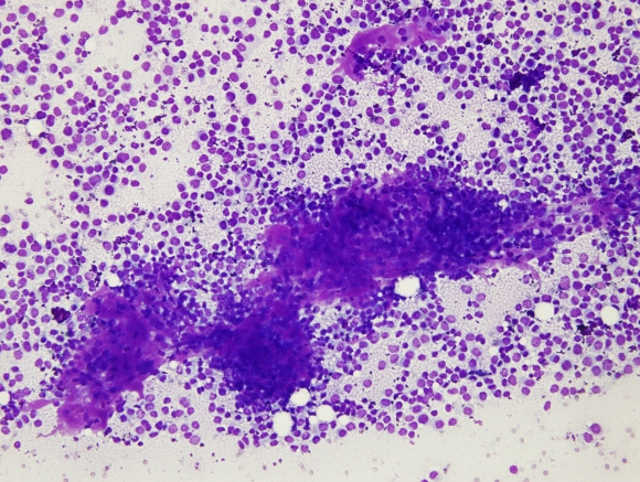 |
| Fig. 1 |
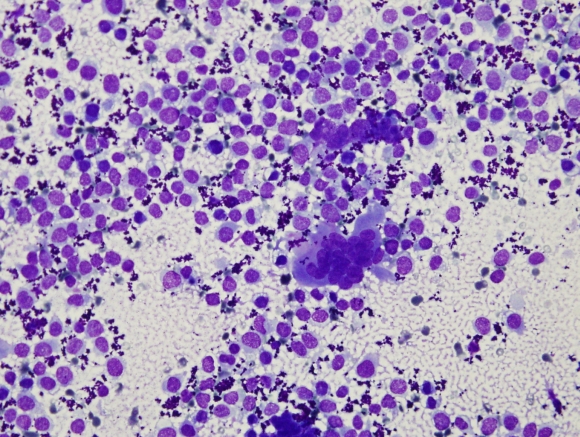 |
| Fig. 2 |
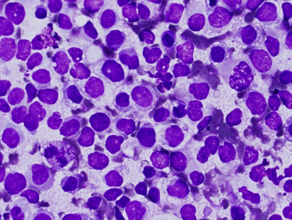 |
| Fig. 3 |
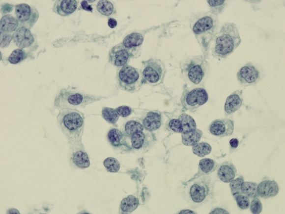 |
| Fig. 4 |
Based on the cytomorphology, the most likely diagnosis of this adnexal mass on FNA is: