September 2010
Aparna Mahajan, MD, Fellow in Cytopathology, Northwestern University, Feinberg School of Medicine, Chicago, IL, U.S.A.
CASE HISTORY:
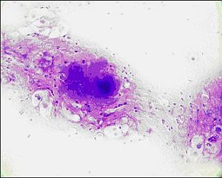
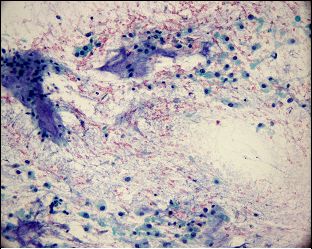
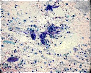
A 50 year old male patient presented with a slow growing sacral mass. Imaging studies revealed a large soft tissue mass measuring 15 cm extending into the bone. Ultrasound guided FNA and core biopsy was performed. FNA smears and touch preparations were made during on site adequacy evaluation/triage by Cytopathology.
 |
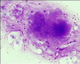 |
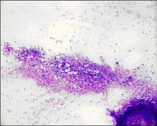
| Fig. 1 FNA Smear, Diff-Quik stain | Fig. 2 FNA Smear, Diff-Quik stain |
 |
 |
| Fig. 3 FNA Smear, Papanicolaou stain | Fig. 4 FNA Smear, Papanicolaou stain |
 |
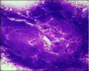 |
| Fig. 5 Touch preparation, Diff-Quik Stain | Fig. 6 Touch preparation, Diff-Quik Stain |
 |
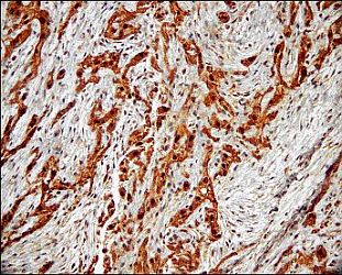 |
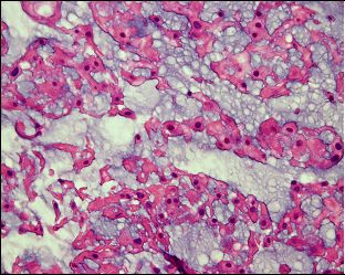
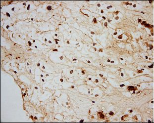
| Fig. 7 Core biopsy H&E Stain | Fig. 8 Core biopsy S100 IHC |
 |
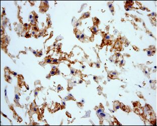 |
| Fig. 9 Core biopsy AE1/AE3-IHC | Fig. 10 Core biopsy EMA-IHC |
Your diagnosis?