November 2012
Author: Alice Laser, MD. Department of Pathology, North Shore/Long Island Jewish Health System, Lake Success, NY, USA
Reviewer: Laura Tabatabai, MD, Department of Pathology, University of California, San Francisco, USA
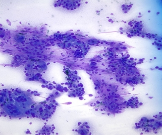
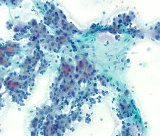
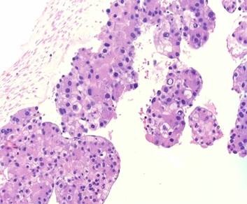
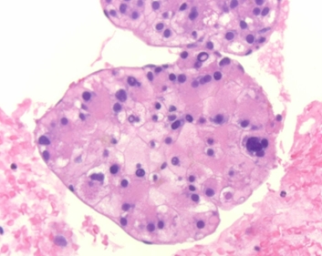
Case History: 92 year old male with elevated liver function tests and found to have an 8.7 x 10 cm mass on ultrasound; the patient has no history of cirrhotic liver disease; ultrasound guided fine needle aspiration biopsy was performed and showed the following:
 |
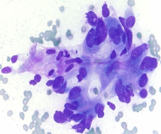 |
| Figure 1: MGG stained smear 10x | Figure 1: MGG stained smear 10x |
 |
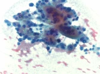 |
| Figure 3: PAP stained smear 10x | Figure 4: PAP stained smear 40x |
 |
 |
| Figure 5: H&E Cell block | Figure 6: H&E Cell block 40x |
Your diagnosis?