March 2018
Author: Angela Chong, MD, Norman Chan, MD, Issam Jajeh, MD.
Dept Anatomical Pathology, Singapore General Hospital, Singapore
Reviewer: ??
The patient, presented with a history of recent cough and a history of treated Pulmonary tuberculosis of uncertain duration . Investigations revealed a mass like consolidation with dilated cystic spaces in the right lower lobe basal segments associated with changes in keeping with mucoid impaction, dilated airways and bronchocele. In view of the previous history of tuberculosis, this was thought to be a reactivation of the disease with endobronchial spread . As the disease did not improve a FNA and core biopsy were performed.
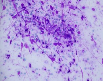 |
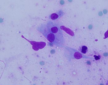
| Fig. 1 low power, DQ stain showing spindled cells with fragile cytoplasm. Background is proteinaceous |
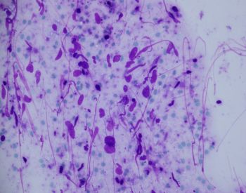 |
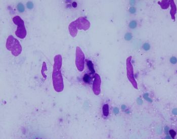
| Fig. 2. low power, DQ stain showing spindled cells. Nuclei are wavy, elongated with an admixture of rounded cells |
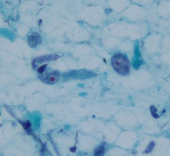 |
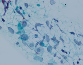
| Fig. 3. PAP stain showing single spindled cells. Nucleoli occasionally rather prominent. May see single or multiple nucleoli |
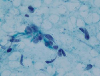 |
| Fig. 4. 4.PAP stain showing similar clustered spindled cells. Nucleoli can be seen clearly with PAP stains |
 |
| Fig. 5. DQ Eloingated and rounded nuclei, some with cytoplasm still present |
 |
| Fig. 6. DQ Elongated nuclei |
 |
| Fig. 7. PAP stain showing cluster of irregular spindle cells |
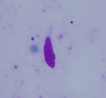 |
| Fig. 8. DQ single cell with fragile cytoplasm |
What is your diagnosis?