June 2014
June 2014
Author: Longwen Chen, MD, PhD, Dept. of Laboratory Medicine and Pathology, Mayo Clinic, Scottsdale, Arizona, USA
Reviewer: Christopher van den Bussche, MD, MIAC, Dept. of Pathology, Johns Hopkins Hospital, Baltimore, MD, USA
Clinical history
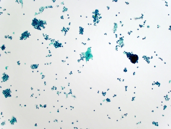
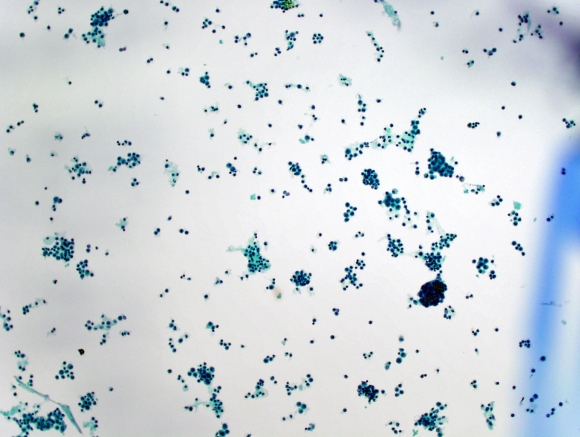
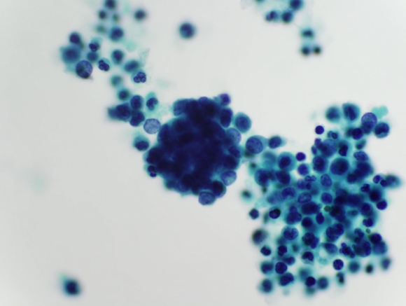
A 63-year-old postmenopausal female presented with a gradual progressive abdominal distension with difficulty in respiration for 2 months. Her serum CA-125 was elevated. Ultrasonography revealed massive ascites and a 2.5 cm mass in the lower abdomen. CT also showed a well-defined pelvic mass measuring 2.5 x 2.2 x1.8 cm. According to the patient’s gynecologist, the patient had a total abdominal hysterectomy and bilateral salpingo-oophorectomy 13 years ago for benign disease. An ascites fluid cytology specimen was obtained.
 |
| Fig. 1 |
 |
| Fig. 2 |
 |
| Fig 3 |
 |
| Fig. 4 |
 |
| Fig. 5 |
Based on the cytomorphology of the ascites fluid (Figures 1-5), what is the most likely diagnosis?