July 2014
July 2014
Author: Zahra Maleki, MD, FCAP, MIAC, Department of Pathology, The Johns Hopkins Hospital, Baltimore, Maryland, USA
Reviewer: Christopher van den Bussche, MD, MIAC, Dept. of Pathology, Johns Hopkins Hospital, Baltimore, MD, USA
Clinical History
A 53 year old woman with a history of lymphocytic thyroiditis was found to have a palpable mass on neck examination. Ultrasound imaging showed a 3.1 cm right thyroid lobe nodule that was heterogeneous with microcalcifications, irregular margins, and positive intranodular flow. Fine needle aspiration biopsy was performed.
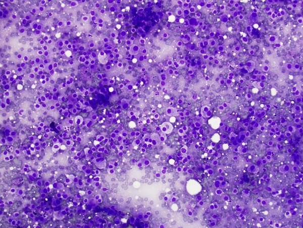
Fig. 1
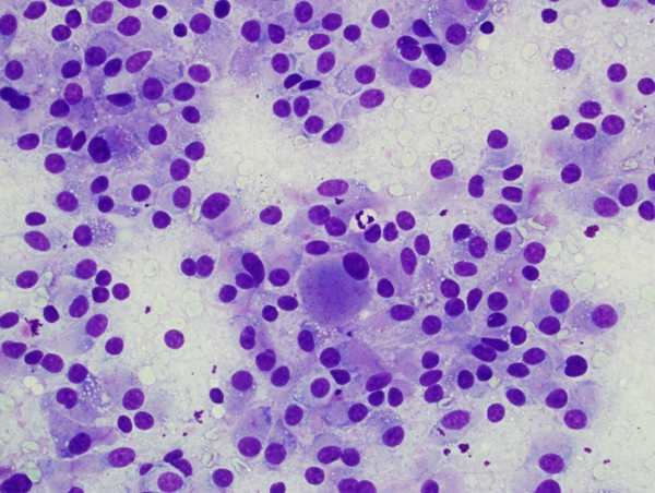
Fig. 2
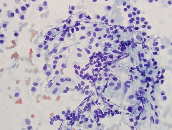
Fig. 3
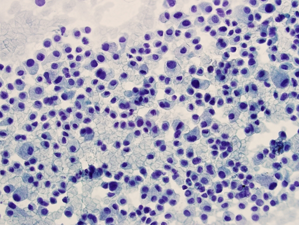
Fig. 4
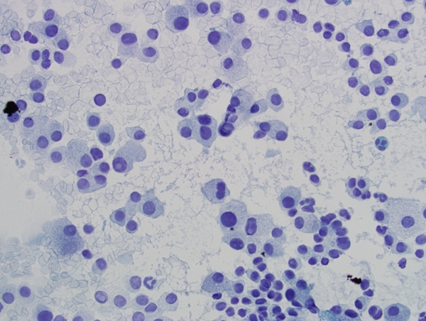
Fig. 5
Based on the cytomorphology of the thyroid nodule FNA what is the most likely diagnosis?