July 2010
Katherine Kasper, MD, Fellow, Department of Cytopathology
Northwestern University, Feinberg School of Medicine, Chicago, IL, USA
CASE HISTORY:
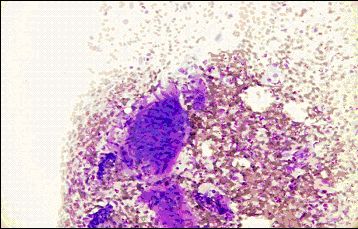
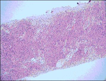
The patient is a 59 year-old male with a history of diabetes and hyperlipidemia who presented with one year of abdominal discomfort, chronic constipation, and increasing abdominal distention. CT scan revealed a large, approximately 25 cm mesenteric mass extending from the upper abdomen to the pelvis. Ultrasound-guided core biopsy was performed. Touch preparations were made during on site adequacy evaluation/triage by Cytopathology.
 |
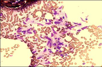 |
| Fig. 1 DQ-stained touch preparations | |
 |
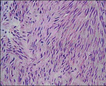 |
| Fig. 2 H&E-stained core biopsy | |
 |
 |
 |
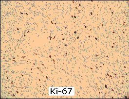 |
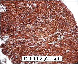
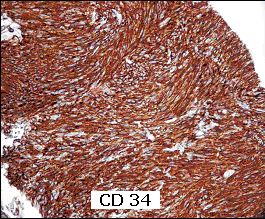
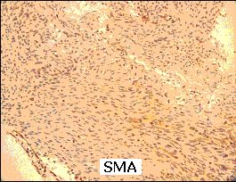
| Fig. 3 Immunohistochemical stains, CD117/c-kit, CD34, smooth muscle actin (SMA), and Ki-67 | |
Your diagnosis?