February 2018
Authors:
Zitong Zhao, Chee Leong Cheng, Jabed Iqbal, Li Yan Khor.
Department of Anatomical Pathology, Division of Pathology, Singapore General Hospital, Singapore.
Reviewer:
Clinical History:
A 30-year-old Malay male patient presented with extensive bilateral cervical lymph node (LN) enlargement for one year. A fine needle aspiration (FNA) biopsy of right level IV cervical LN was performed for a diagnostic work-up.
Cytopathology:
FNA smears showed a cellular yield of large atypical cells in a polymorphous background (Figure 1). The background showed maturing lymphocytes, plasma cells, neutrophils and eosinophils (Figure 2). The large atypical cells had mono-, bi- and multilobate bizarre nuclei with clumped chromatin and scanty cytoplasm (Figure 3), which were rare in the cell block (Figure 4).
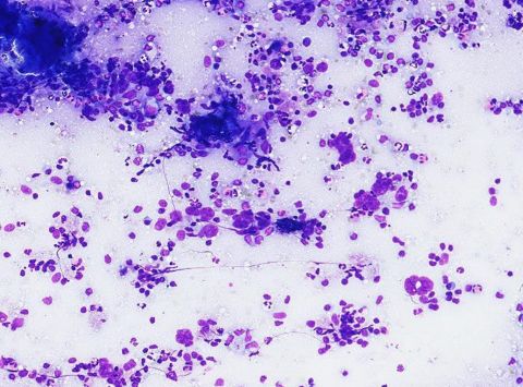 |
| Fig. 1 . Cervical lymph node, Diff Quik x 10 |
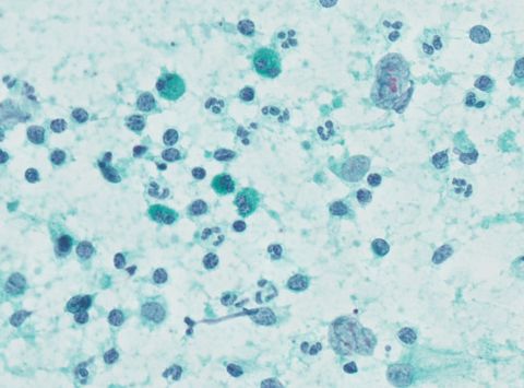 |
| Fig. 2. Cervical lymph node, Pap x 40 |
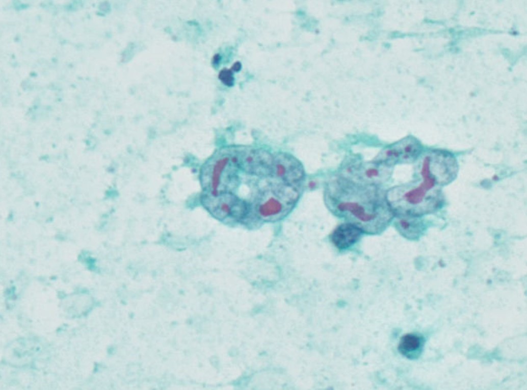 |
| Fig. 3. Cervical lymph node, Pap x 60 |
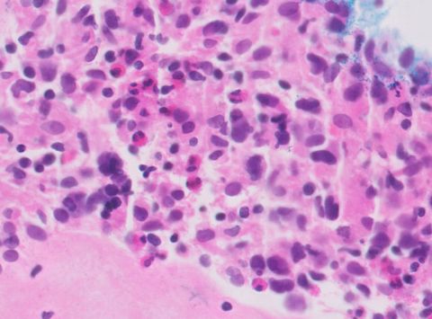 |
| Fig. 4. Cervical lymph node, cell block, H&E x 40 |
What is your diagnosis?