August 2010
Walid H. Khalbuss, MD, PhD, FIAC & Amer Heider, MD
Dept of Pathology, University of Pittsburgh Medical Center (UMPC), Pittsburgh, PA, USA
CASE HISTORY:
The patient is a 33-year old female with no history of malignancy who presents with (10 cm) liver mass. CT-guided Liver FNA was performed. 5 images are provided.
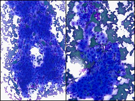 |
Fig. 1 DQ-stained smears x100 left x200 right |
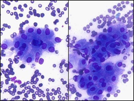 |
Fig. 2 DQ stained smears x400 |
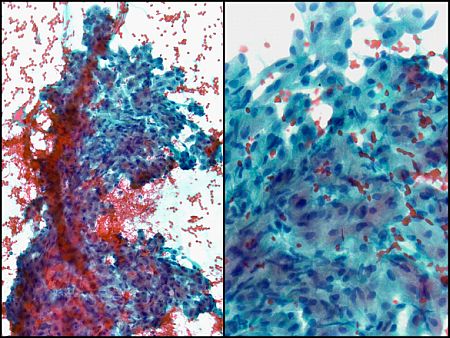 |
Fig 3 Pap-stained smear x100 left x200 right |
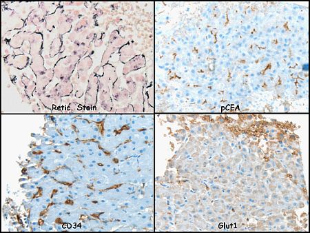 |
Fig. 4 Ancillary studies of Reticulum stain, pCEA, CD34 and Glut 1 |
Your diagnosis?