October 2015
Authors: Immacolata Cozzolino MD, Ph.D1, Chiara Mignogna MD,Ph.D2, Pio Zeppa MD,Ph.D
Departments of 1Public Health, University of Naples “Federico II”, 2Pathology University “Magna Graecia” of Catanzaro, Medicine and Surgery, University of Salerno, Italy.
Corresponding Author: Prof. Pio Zeppa, Azienda Ospedaliera Universitaria San Giovanni di Dio e Ruggi d’Aragona, Salerno, Italy
Reviewer: Christopher J. VandenBussche MD PhD, The Johns Hopkins University School of Medicine, Baltimore, Maryland, USA
CASE HISTORY
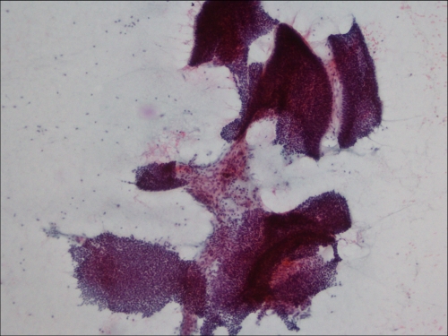
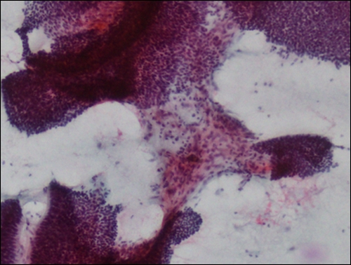
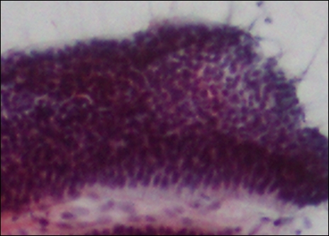
A 60-year-old woman, with no significant previous medical history, showed a painless swelling in the left jaw for 2 years. X-rays showed a 24 mm, lobulated osteolytic lesion with pushing borders and cortical thinning (fig. 1). Serologic parameters were within normal ranges and the total body CT scan did not show any other pathology. FNA was performed with a transmucosal approach without radiological guidance. Diff-Quik and Papanicolaou stained conventional smears were prepared (figs. 2-7)
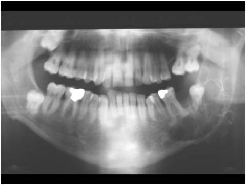 |
| Fig. 1 |
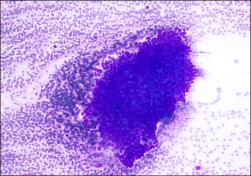 |
| Fig. 2 |
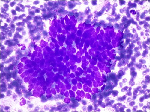 |
| Fig. 3 |
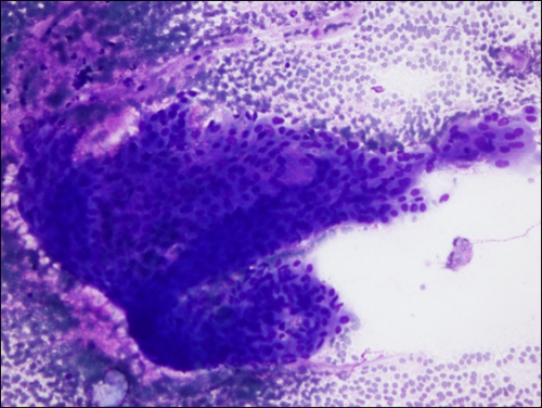 |
| Fig. 4 |
 |
| Fig. 5 |
 |
| Fig. 6 |
 |
| Fig. 7 |
What is your diagnosis?