October 2010
Katherine Kasper, MD
Fellow, Dept. of Cytopathology
Northwestern University, Feinberg School of Medicine, Chicago, IL, USA
CASE HISTORY:
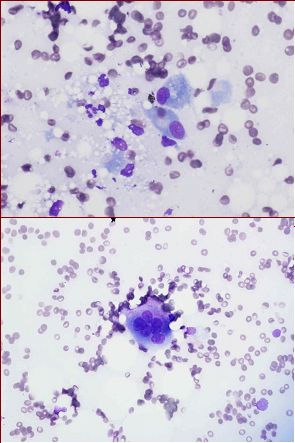
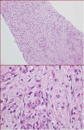
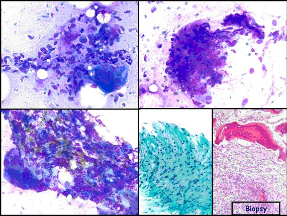
The patient is a 46 year-old male with a history of recent hemorrhagic stroke undergoing rehabilitation, who was found to have right medial thigh swelling. CT imaging revealed an approximately 4 cm soft tissue lesion, suspected radiographically to be myonecrosis. Ultrasound-guided core biopsy was performed. Touch preparations were made during on site adequacy evaluation/triage by Cytopathology.
 |
 |
| Fig. 1. DQ stained touch preparations | Fig. 2. H & E stained core biopsy |
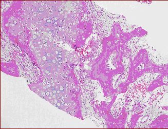
| Fig. 3. H & E stained core biopsy | Fig. 4. H & E stained core biopsy |
 |
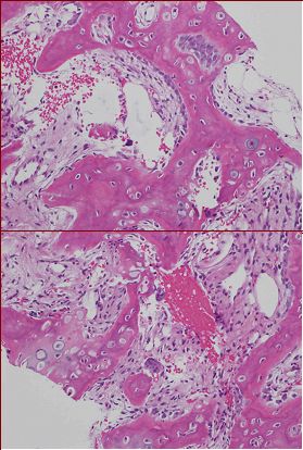 |
| Fig. 5. FNA case and surgical excision of Myositis ossificans showing zonal phenomenon with a mixed cytomorphological patterns including myxoid (upper right); edematous (upper left); bone tissue (lower left), inflammatory cells; hypocellular areas, and hypercellular areas. The proliferating cells are mixture of spindle, stellate fibroblasts and giant cells. (Courtsey: Dr Walid Khalbuss) | |
 |
|
Your diagnosis?