May 2013
May 2013
Authors: Jennifer M. Harness BS and Sara E. Monaco MD; Department of Pathology, University of Pittsburgh Medical Center, Pittsburgh, PA USA.
Reviewer: Walid E. Khalbuss, MD PhD FIAC, Department of Pathology, University of Pittsburgh Medical Center, Pittsburgh, PA USA.
Clinical History: The patient is a 29 year old female who presented with menorrhagia. She was evaluated with transvaginal pelvic ultrasound and incidentally noted to have a hyperechoic density in the mid-lateral pole of the right kidney. A CT-scan performed 6 months later revealed a round, hyperdense, enhancing, well-circumscribed lesion that measured 3.1 cm in greatest dimension. Thus, a CT-guided fine needle aspiration of the right kidney mass was performed with rapid on-site evaluation. Images (Figures 1-4 are provided).
 |
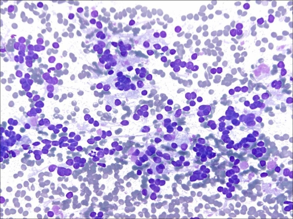
| Fig. 1 CT-guided FNA of renal mass (DQ stain, original magnification x400). |
 |
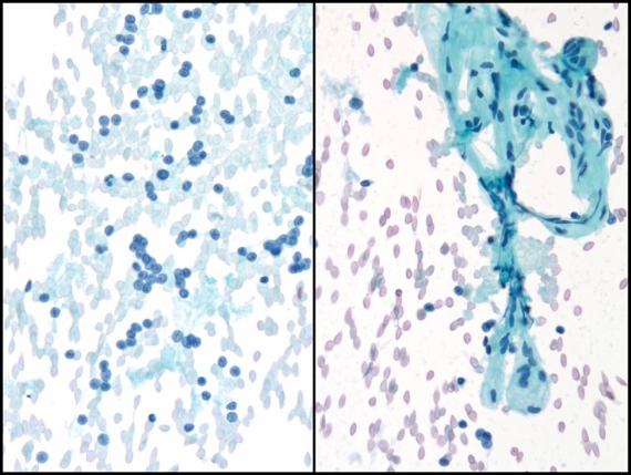
| Fig. 2 CT-guided FNA of renal mass (Pap stain, original magnification x400) |
 |
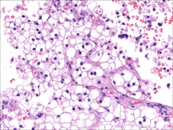
| Fig. 3. CT-guided FNA of renal mass (cell block, H&E stain, original magnification x400). |
 |
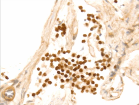
| Fig. 4. : CT-guided FNA of renal mass (TFE3 immunostain, original magnification x400). |
What is the best diagnosis for this specimen?