July 2011
Mohamed Aziz, MD, Seema Lale, MD, Patricia Wasserman, MD, North Shore – Long Island Jewish Pathology and Lab Medicine, Lake Success, New York, USA
CASE HISTORY:
A 66 year old female was referred to the hospital by her cardiologist to manage a bloody pericardial effusion. The patient reported to her cardiologist with medical cardiac symptoms. Pericardial tap produced 90 ml of thick bloody fluid. No significant history was known at the time of presentation. Cytology smears and cell block preparation were prepared.
|
|
 |
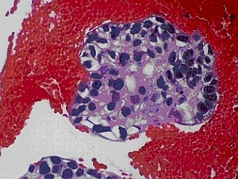
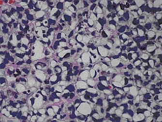
| Fig. 1A – H & E stained cell block (x 40) | Fig. 1B – H & E stained cell block (x 40) |
 |
 |
| Fig. 2A – H & E stained cell block (x 20) | Fig. 2B – H & E stained cell block(x 40) |
 |
 |
| Fig. 3 Ancillary studies of CK7, CK20, EMA, CK5/6, Mucin and PAS |
Your diagnosis?
