January 2016
Authors:
Preetha Alath MBBS, MD, Department of Pathology, Kuwait Cancer Control Center, Kuwait
Kusum Kapila, MD, FAMS, FIAC, FRCPath, Professor and Head of Cytopathology Unit, Department of Pathology, Faculty of Medicine, Kuwait University
Reviewer: Christopher J. VandenBussche, MD, PhD, The John Hopkins University School of Medicine, Baltimore, Maryland, U.S.A.
Clinical History
A 44 year-old woman presented with a nodule in the upper outer quadrant of the left breast. Sonomammography showed a well-defined, heterogenous, solid nodule with posterior acoustic enhancement, lateral marginal shadowing and a vascular core (BIRADS IV) in the upper outer quadrant of the left breast. FNA was performed.
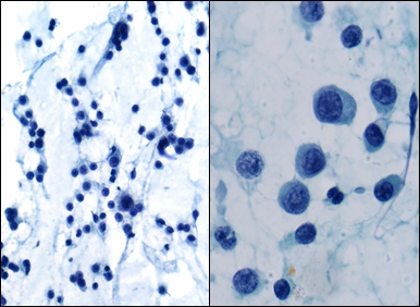
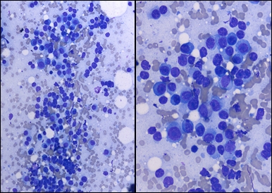
Figure 1 and Figure 2 are Papanicolaou and MGG stained smears from the breast mass.
 |
| Fig. 1 |
 |
| Fig. 2 |
Based on cytomorphology (Figures 1 and 2) what is the most likely diagnosis?