January 2013
Author: Richard Cantley, MD, Cytopathology Fellow,
Northwestern University, Feinberg School of Medicine,Chicago, IL 60611
Reviewer: Walid Khalbuss, MD, FIAC
A 59 year old female presented with right upper quadrant abdominal pain. A 4.9 cm unilocular cystic hepatic mass was discovered on MRI. Aspiration of the cyst fluid was performed. Four cytospin slides (2 Diff Quik stained and 2 Papanicolaou stained) and one Hematoxylin and Eosin (H&E)-stained cell block slide were prepared. Immunohistochemical stains performed for ER and PAX-8 on cell block material were negative.
 |
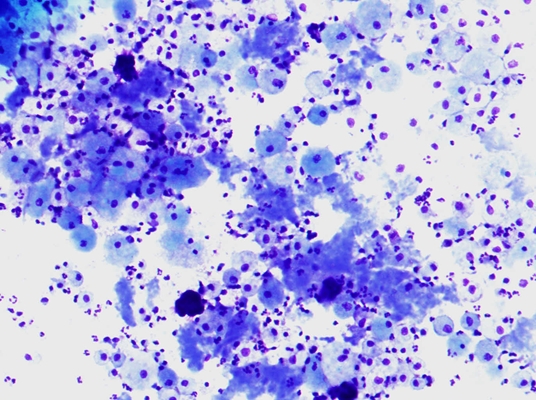
| Image 1, Diff-Quik Stain, 200x |
 |
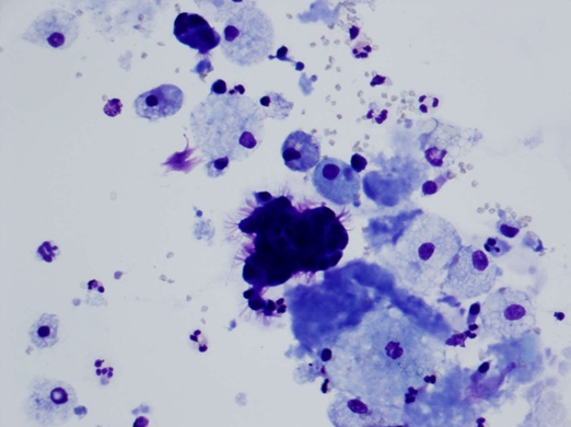
| Image 2, Diff-Quik Stain 400x |
 |
| Image 3, Diff-Quik Stain 400x |
 |
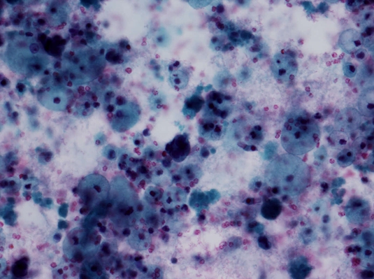
| Image 4, Papanicolaou Stain 400x |
 |
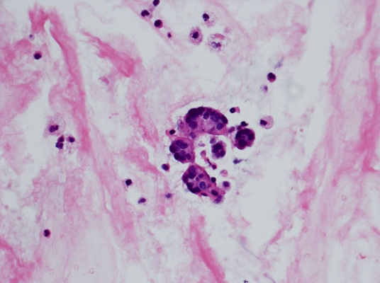
| Image 5, Cell Block, H&E stain 400x |
Which of the following is the correct diagnosis?