December 2011
Antonio D’Antonio MD, Pio Zeppa MD
Dipartimento di Anatomia Patologica, Azienda Ospedaliera Universitaria, San Giovanni di Dio e Ruggi D’Aragona, Salerno Italy.
Case history:
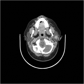
An 8-year-old boy complained of a rapidly growing, painless, nodule in the right parotid region. An ultrasonographic evaluation revealed a 2.7 x 2.5 cm, hypoechogenic mass within the right parotid gland with sharp, well-defined borders and without an evident capsule. A magnetic resonance imaging (MRI) confirmed the presence of an intraparotid well-circumscribed, solid, unencapsulated nodule; no other masses or lymph nodes were detected. (Fig.1).
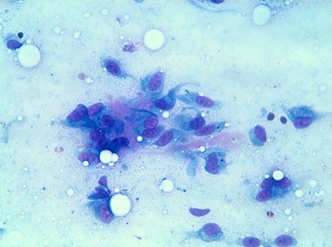
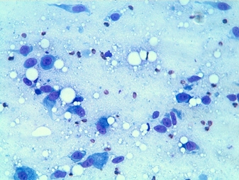
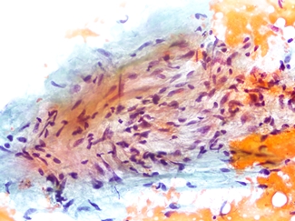
A fine-needle aspiration (FNA) was performed under ultrasound control and two air-dried smears were Diff-Quik stained and immediately evaluated. A second pass was then performed and additional smears were fixed in 95% alcohol and stained with Papanicolaou stain.
 |
| Fig. 1.MRI showing a well-circumscribed, 27 mm, solid nodule in the right parotid. Note the thin rim of the gland in the posterior side |
 |
| Fig. 2. Diff-Quik stain, 430X. |
 |
| Fig. 3. Diff-Quik stain, 430X |
 |
| Fig. 4. Papanicolau stain. 430X |
Your diagnosis?