December 2010
Manon Auger, MD, FRCP(C), MIAC
Director, Cytopathology Laboratory, McGill University Health Center
Associate Professor, Department of Pathology, McGill University
Montreal, PQ, Canada
CASE HISTORY:
Fine needle aspirate of a 2 cm nodule in the right lobe of the thyroid in a 69 year old woman.
Cytomorphology of the case: (Fig.1-9)
The smears are very cellular consisting of a monotonous population of Hurthle cells characterized by abundant granular cytoplasm, a round eccentrically located nucleus, and a prominent nucleolus. The Hurthle cells are arranged predominantly in a syncytial or trabecular arrangement; many single Hurthle cells are also noted. Some of the cells are seen in close connection to capillaries (Fig.5). Macrophages, indicative of cystic degeneration, are seen in the background.
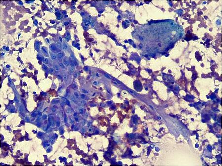 |
1-Diff-Quik stain, 200X |
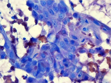 |
2-Diff-Quik stain, 400X |
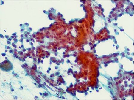 |
3- Papanicolaou stain, 200X |
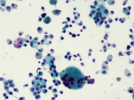 |
4-Papanicolaou stain, 200X |
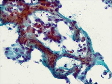 |
5-Papanicolaou stain, 200X |
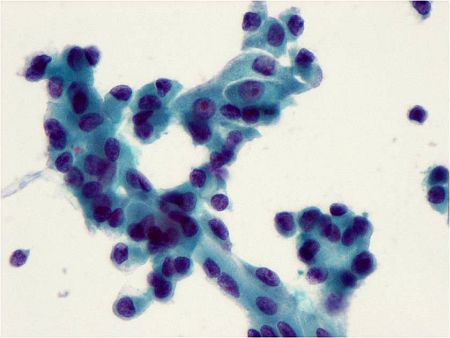 |
6-Papanicolaou stain, 400X |
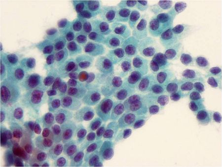 |
7-Papanicolaou stain, 400X |
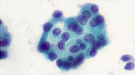 |
8-Papanicolaou stain, 600X |
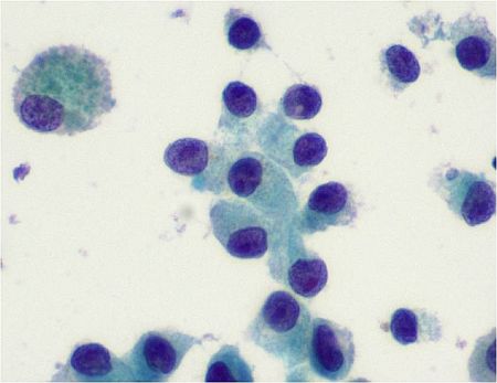 |
9-Papanicolaou stain, 600X |
Your diagnosis?