August 2013
August 2013
Authors: Nicole Bures MD and Sara E. Monaco MD; University of Pittsburgh; Pittsburgh, PA; USA
Reviewer: Claire W. Michael, M.D. Case Western Reserve University, Cleveland, Ohio, USA
Case History:
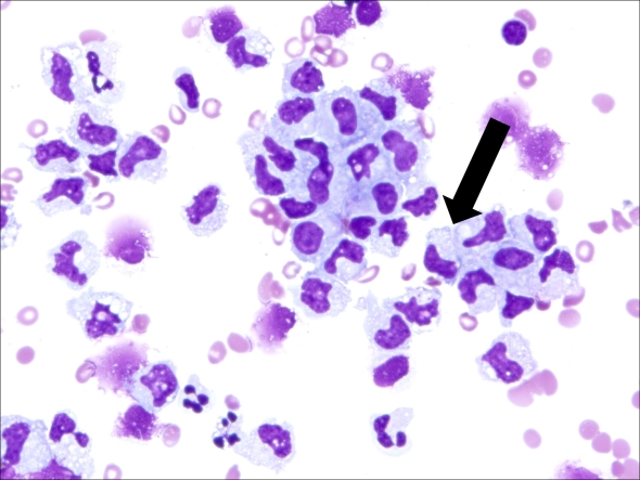
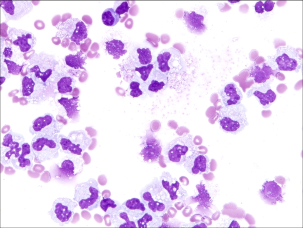
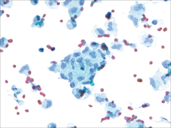
43-year-old man with end-stage renal disease presents with ascites. The patient has a history of thrombophilia, including a deep vein thrombus, pulmonary emboli, and splenic thrombi. He is also HIV positive and has had a history of granulomatous lymphadenitis involving pelvic lymph nodes. The ascites fluid was sent for cytological evaluation and flow cytometry. The cytology laboratory received 1000 cc of cloudy yellow fluid. Cytospins and a cell block were prepared. Representative images of the cytospin slides are seen in Figures 1-3. Flow cytometry performed on the specimen revealed heterogeneous T-cells and polytypic B-cells.
 |
| Figure 1 (Cytospin, DQ stain, original magnification x 400). |
 |
| Figure 2 (Cytospin, DQ stain, original magnification x 400) |
 |
| Figure 3 (Cytospin, Pap stain, original magnification x 400). |
What is the best diagnosis for this patient?