March 2012
Authors: Juan Xing, MD1, Walid E. Khalbuss, MD PhD FIAC2, Liron Pantanowitz, MD, MIAC2
1. Department of Pathology, Drexel University College of Medicine, Philadelphia, PA, USA
2. Department of Pathology, University of Pittsburgh Medical Center, Pittsburgh, PA, USA
CASE HISTORY:
A 59 year old male presented to an outside institute with a slowly growing left-sided chest wall mass which was surgically resected. Six years later he recurred with a new chest wall mass and masses in his left lung and abdomen.. Now his chest wall mass has doubled in size. A FNA with air-dried Diff Quik stain and a cell block of the recurrent chest wall mass was performed.
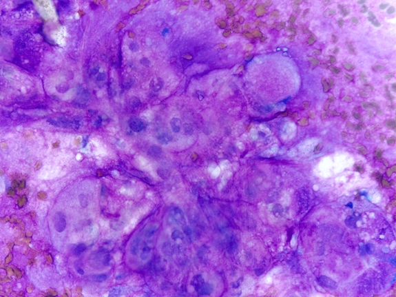 |
Fig. 1 FNA direct smear of the recurrent left chest wall mass (DQ stain, Mag 40x)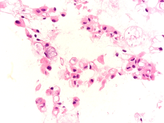 |
| Fig. 2 Cell block preparation showing tumor cells of the recurrent left chest wall mass (H&E stain; Mag 40x) |
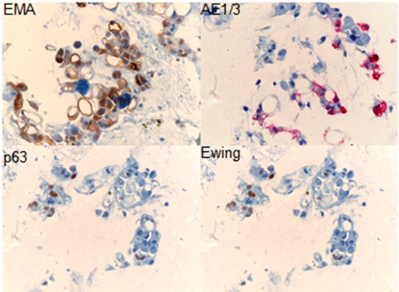 |
| Fig. 3 Several immunostains performed on cell block material of the recurrent chest wall mass are shown. |
What is the best interpretation for this case?