June 2011
Nidhi Aggarwal, MD and Walid E. Khalbuss, MD, PhD, FIAC. Dept. of Pathology, University of Pittsburgh Medical Center (UMPC), Pittsburgh, PA, USA
CASE HISTORY:
69-year-old male with no history of malignancy presented with enlarged mesenteric lymph nodes the largest measuring 3.4 cm. US-Guided FNA was performed. (Images 1-3).
ICD-O Site: Lymph node FNA biopsy cytology
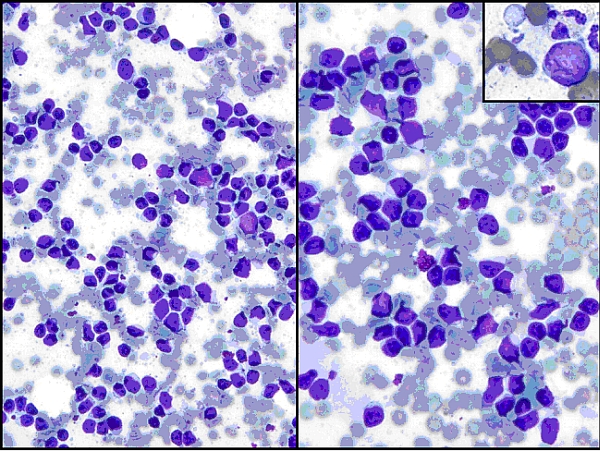 |
| Image 1 (Wright Giemsa) |
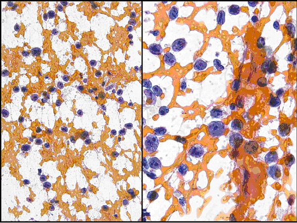 |
| Image 2 (Papanicolau stain) |
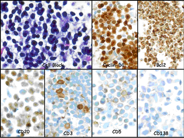 |
| Image 3 (Immunohistochemistry) |
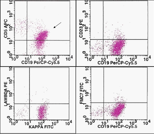 |
| Image 4 (Flow cytometry) |
|
|
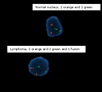
| Image 5 (Interphase Fluorescence In-Situ Hybridization Dual-color dual-fusion probe |
The most likely diagnosis of this lymph node specimen is?