July 2013
July 2013
Authors: Rebecca J Leeman-Neill, MD PhD and Walid E. Khalbuss, MD PhD FIAC, University of Pittsburgh Medical Center (UPMC), Pennsylvania, USA
Reviewer: Ritu Nayar; MD; Northwestern University; Chicago, IL, USA.
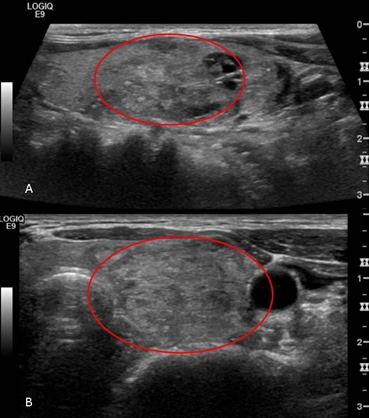
Clinical History: The patient is a 58 year old woman with a 3.4 x 2.3 x 1.3 cm solid nodule with some small cystic spaces occupying the mid and lower left pole of the thyroid, as seen on ultrasound imaging (Figure 1A: longitudinal plane, 1B: transverse plane). The patient has noted an increase in size of this nodule and associated dysphagia. TSH and T3 are within normal limits. The patient also has a history of low-grade lymphoma diagnosed 8 years ago, after presenting with cervical lymphadenopathy, for which she has received intravenous immunoglobulin (IVIG) treatments.
FNA of thyroid nodule was performed and 5 images are provided
 |
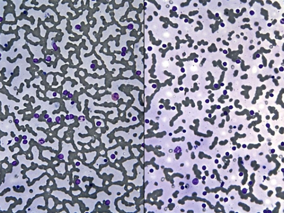 |
| Image 2: DQ-stained smears (x100) |
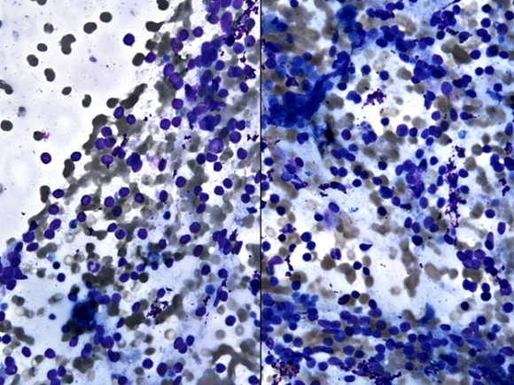 |
| Image 3: DQ-stained smears (x200) |
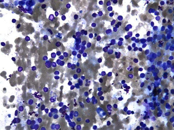 |
| Image 4: DQ-stained smears (x400) |
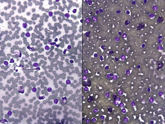 |
| Image 5: DQ-stained smears (x400) |
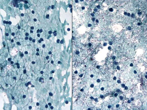 |
| IMAGE 6 : Pap-stained smear (x400) |
What is the most likely diagnosis of this case?