January 2011
Tee Lang, M.D. and Walid E. Khalbuss, M.D., Ph.D., FIAC.
University of Pittsburgh Medical Center (UPMC), Pennsylvania, USA
CASE HISTORY:
The patient is a 71-year old female who has recent CT and MRI suggestive of large renal mass englobing the left kidney and retroperitoneal lymph node and a lytic bone lesion in the L5 of vertebral body. FNA of L5 vertebral body was performed and 4 images are provided:
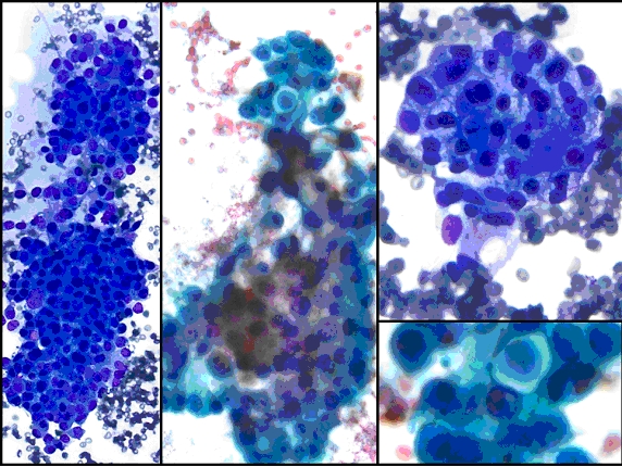 |
| Image 1: DQ-stained smears (x100, left), x400 (right upper). And Pap-stained smear (x200, middle and x600 lower right) |
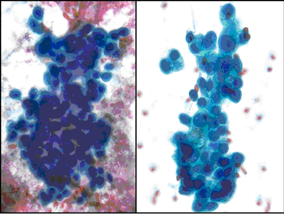 |
| Image 2: Pap-stained smear, x200 (left), x400 (right). |
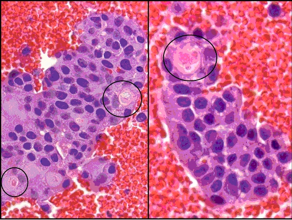 |
| Image 3: Cell block (H&E stain, x400 left and 600 right) |
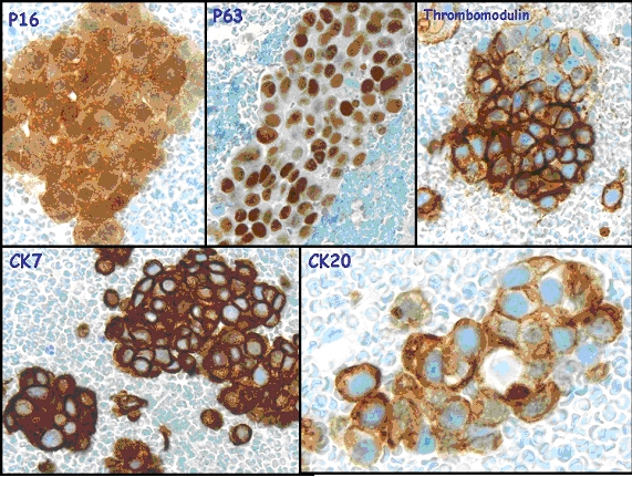 |
| Image 4: Ancillary studies of Ck7, CK20, p16, p63, and thrombomodulin. |
Your diagnosis?