February 2014
February 2014
Author:
Longwen Chen, MD, PhD, Dept. of Laboratory Medicine and Pathology, Mayo Clinic, Scottsdale, Arizona, USA
Reviewer: Andrew S. Field, MB, BS, FRCPA, Dept. of Pathology, St. Vincent’s Hospital, Sydney, Australia
Clinical history
A 27-year-old female was diagnosed with a mass in the pancreatic body that was detected incidentally by abdominal ultrasonography during a general examination. The mass is 4.2 cm in maximum dimension. The tumor was well-defined and hypoechoic on EUS imaging. EUS-FNA was performed via a transgastric approach without complications.
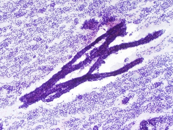 |
| Fig. 1 |
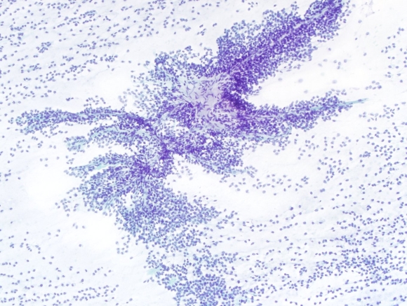 |
| Fig. 2 |
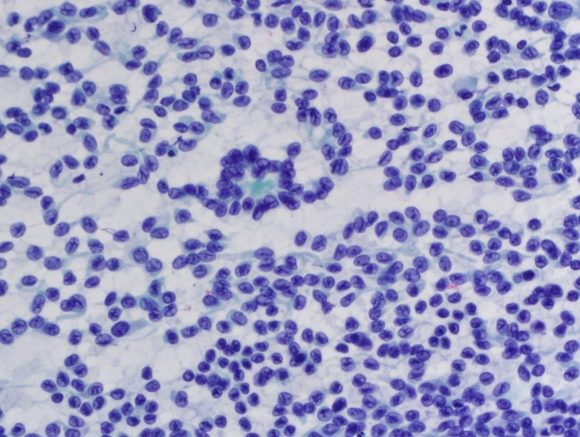 |
| Fig. 3 |
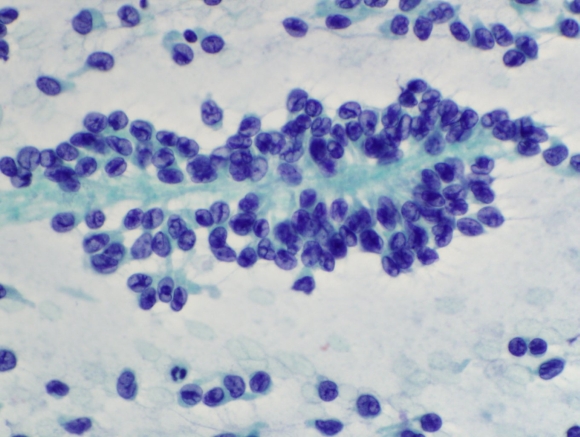 |
| Fig. 4 |
Based on the cytomorphology of the EUS-FNA, what is the most likely immunoprofile of this tumor?