December 2012
Authors: Walid E. Khalbuss, MD PhD FIAC; and Shveta Hooda MD, Department of Pathology, University of Pittsburgh Medical Center (UPMC), Pittsburgh, PA, USA.
Reviewer: Manon Auger, M.D, FRCP(C), Department of Pathology, McGill University and McGill University Health Center, Montreal, PQ, Canada
Case presentation:
A 81 year old female with past medical history of hypertension, hypothyroidism, hyperlipidemia and chronic kidney disease presented with abdominal pain and vomiting after eating food. Physical examination elicited pain scaled around 3-4/10 localized in the epigastric area. The patient was found to have paroxysmal atrial fibrillation with rapid ventricular response and a left pleural effusion (Figure 1). Pleural fluid (50 ML of thick, light brown opaque fluid) was submitted for cytological evaluation (Figures 2-5).
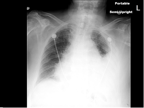 |
| Fig. 1 (Chest X-ray, semi upright with left pleural effusion) |
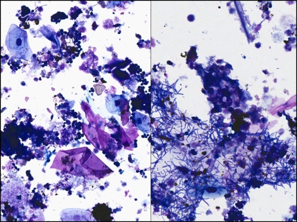 |
| Fig. 2 (DQ stain, high magnification) |
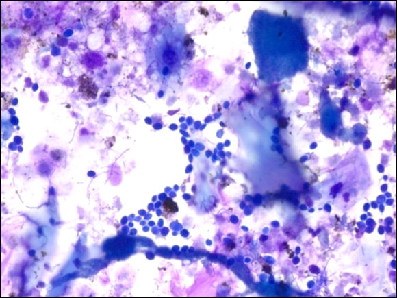 |
| Fig. 3 (DQ stain, high magnification) |
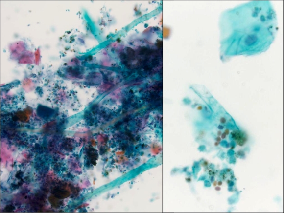 |
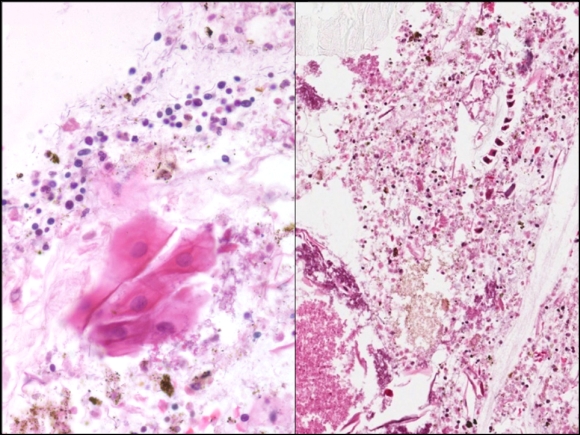
| Fig. 4 (Pap stain, low magnification, left, and high magnification, right |
 |
| Fig. 5 (Cell block, H&E stain, high magnification) |
Which of the following best describes depicted lesion?